



For molecular modelers, aligning protein structures can often feel like a tedious and complex task. Whether you’re identifying conserved residues, comparing conformations, or preparing homology models, finding a reliable workflow is essential. SAMSON’s Protein Aligner provides an integrated solution to align protein sequences and superimpose structures, all in a streamlined and user-friendly interface.
Why Align Proteins?
Protein alignment allows researchers to:
- Identify conserved residues critical for function or ligand binding.
- Compare protein conformations across species or mutations.
- Create more accurate homology models for further molecular design workflows.
These capabilities make protein alignment a cornerstone of molecular modeling workflows, whether you’re studying evolutionary relationships or designing novel molecules.
Getting Started: Preparing Your System
To begin using Protein Aligner in SAMSON, follow these steps to ensure a clean and optimized configuration:
- Fetch or load the protein structures you wish to align. For example, you can align two hemoglobins,
1DLWand1RTX, by opening Home > Fetch and inputting the identifiers. - If your proteins include unnecessary solvent or ligands, utilize SAMSON’s Protein Preparation & Validation tools to clean up the data.
- Enable the Protein Aligner extension by navigating to Home > Align
 .
.

Sequence Alignment
Sequence alignment is the first step toward comparing proteins. SAMSON offers two modes:
- Align sequences (by structure): Aligns one full model to another.
- Align sequences (by chain): Aligns individual chains, an option particularly useful for oligomeric proteins.
Once you’ve selected the appropriate mode, click on Align sequences. Below, you can see a sample alignment comparing two hemoglobin sequences, along with options to highlight conserved residues and amino acid properties like polarity:

Residue colors reflect their properties, and if a residue matches multiple selected properties, its color shows blended shades for easier interpretation.
Structural Superposition
Aligning protein structures in SAMSON is just as intuitive. For whole-protein alignment:
- Ensure no residues are selected.
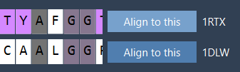
- Click Align to this on the first protein model’s row in the Protein Aligner interface.
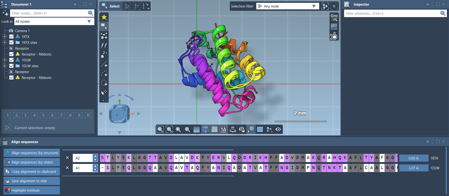
The RMSD (root mean square deviation) value between the two structures will be displayed, indicating their alignment accuracy. For example:
Visualizations in SAMSON can enhance your alignment analysis further. Adding secondary structure representations (e.g., ribbons) can differentiate the aligned proteins more clearly. Adjust these via Visualization > Visual model > Ribbons for individual structures:
Region-Specific Alignment
Sometimes, focusing on specific regions of a protein is more relevant than aligning entire structures. SAMSON allows you to align selected regions seamlessly:
- Select the residues for alignment, such as the first 20 residues in both sequences.
- Click the alignment button next to the selection to superimpose only the selected regions.
Below, you can see an example showing region-specific alignment for two alpha-helices:

Next Steps
Once your alignments are complete, you can:
- Export the alignment for homology modeling workflows.
- Overlay conserved residues onto ligand-binding sites for molecular design.
- Repeat alignments with additional chains or proteins to expand your analysis.
To dive deeper into these workflows, refer to the complete documentation at SAMSON Protein Aligner Documentation.
Note: SAMSON and all SAMSON Extensions are free for non-commercial use. Download SAMSON at https://www.samson-connect.net.