



A common frustration in molecular modeling is having two structures that represent the states you want to connect, but do not line up neatly as endpoints for a transition-path calculation. They may differ in residue count or structure details. A direct interpolation can then be difficult to set up—or can produce a path that does not represent the same molecular system at both ends.
The SARS-CoV-2 spike opening example offers a useful case study. It connects the closed structure, PDB 6VXX, with the open structure, PDB 6VYB. The goal is to explore the change from the down state to the up state, in which the receptor-binding region can recognize ACE2. The important modeling lesson is not simply that the protein moves: it is how the workflow handles endpoints that differ in residue count.
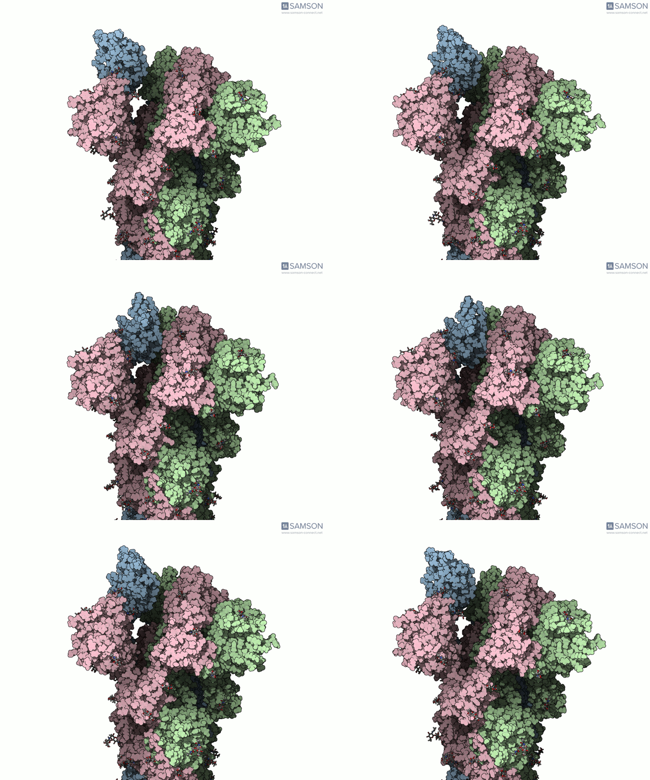
Why the endpoint mismatch matters
A transition path is only useful when its conformations can be interpreted in relation to the structures used to define it. In this example, 6VXX and 6VYB contain different numbers of residues. The ARAP Interpolation Path module generates a path for the structure used as its starting conformation, so simply choosing the two experimental structures as endpoints does not automatically produce a path represented consistently by both structures.
The workflow addresses this by using an intermediate conformation. First, the structures are prepared: bond orders in sugars are corrected, then hydrogens are added and minimization is performed. The open structure is used as the starting conformation and the closed structure as the goal for an initial ARAP interpolation. Because that path corresponds to the starting structure, a closed-like conformation is taken from the path and minimized. That conformation can then serve as a compatible goal for a second interpolation.
This is a practical strategy to consider when endpoint structures are not directly comparable: use the initial path to construct a suitable endpoint in the representation of the starting structure, then generate the path again. It is not a universal fix for every mismatch. The right approach depends on what differs between the structures and which atoms or residues need to be represented in the resulting path.
From an initial path to a refined one
ARAP supplies an interpolated path between the selected conformations. The example then uses the P-NEB (Parallel Nudged Elastic Band) module to improve the path. The documented computation took less than half a minute for the ARAP path and about 15 minutes for the P-NEB step on a laptop. These are timings from this particular example, not performance guarantees for other systems.
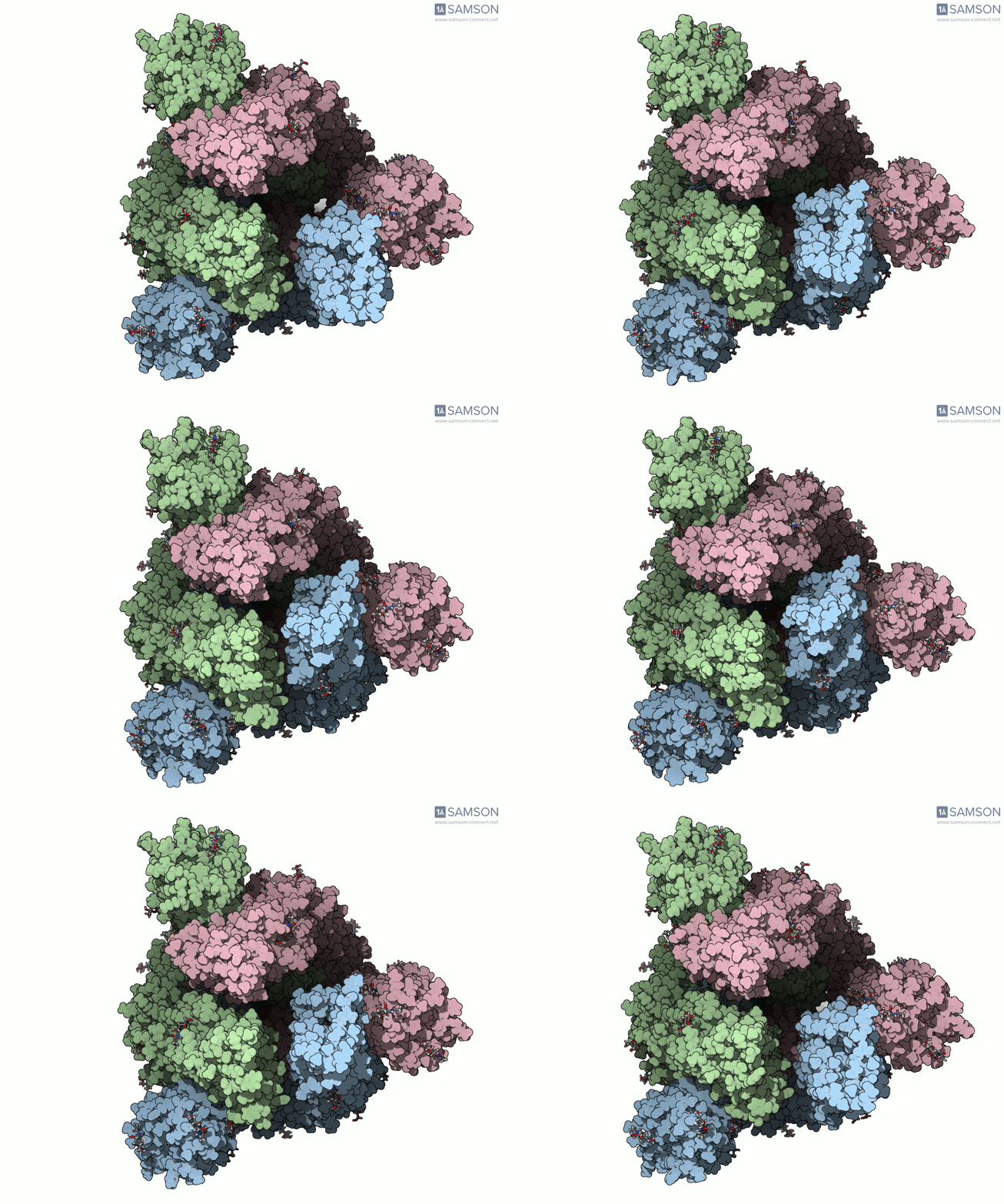
Viewing the motion from multiple angles helps make the structural change easier to interpret. The spike is a trimer, and the open state exposes a receptor-binding region that can interact with ACE2. The animation is a visual representation of a computed path; it should not be mistaken for a recording of the protein moving in real time.
Use the trajectory with appropriate caution
The downloadable files include a set of PDB trajectory files, a single PDB trajectory, and a SAMSON-format file. The SAMSON file brings together the spike structure, open and closed conformations, and paths associated with ARAP and P-NEB. This can make it convenient to inspect the structures and animate the path in the same document.
Crucially, the trajectory is provided as is and has not been experimentally verified. Treat it as an illustrative computed motion, not as proof of a biological mechanism or a validated pathway. If you plan to use it in further calculations, check whether additional post-processing or validation is needed for your specific research question. The structures, animations, images, and trajectories are released under the CC BY 4.0 license.
For the complete walkthrough and the trajectory downloads, see the original documentation page.
SAMSON and all SAMSON Extensions are free for non-commercial use. Get SAMSON at samson-connect.net.