



Two proteins can be related yet look poorly matched when their full structures are superimposed. That does not necessarily mean there is no useful similarity: the region you care about may align well even when other parts differ. For molecular modelers comparing homologs, mutants, or proteins from different organisms, separating sequence alignment from structural superposition—and knowing when to focus on a region—can make the comparison more informative.
This workflow uses hemoglobins 1DLW and 1RTX as an example. The same steps can be applied to other proteins you have loaded in SAMSON.
Start with comparable structures
Fetch or open the proteins before starting the alignment. In SAMSON, use Home > Fetch, enter 1DLW 1RTX in the PDB or PDB (mmCIF) field, and load the structures. If the models contain solvent, ligands, or alternate locations that should not be part of the comparison, prepare them first with Home > Prepare.

Then open Protein Aligner from Home > Align. Before interpreting a structural fit, generate a sequence alignment. This lets you inspect corresponding residues rather than treating the proteins as undifferentiated shapes.
Choose the alignment level that matches your system
Protein Aligner offers two sequence-alignment modes. Align sequences (by structure) aligns one model to another, per structural model. Align sequences (by chain) aligns individual chains, which can be useful when a model contains multiple chains, such as in an oligomer. The right choice depends on whether your comparison is between whole models or between particular chains.

Sequence similarity is only one view of the comparison. You can highlight amino-acid properties such as similarity and polarity to make conserved positions and property differences easier to inspect. A residue that matches more than one selected property is shown with a blended color. This visual check can help you connect an alignment column to the chemical character of its residues.

Use residue selection to connect the alignment to the structure
Hover over a residue to see its name and ID and highlight it in the viewport. Clicking a residue selects it and its aligned partners. You can extend the selection across consecutive residues with Shift, select non-consecutive residues with Ctrl on Windows or Linux or Cmd on macOS, and use Alt to remove residues from the selection. Selecting a residue in the Document view or viewport also highlights it and its aligned partners in Protein Aligner.
This connection between sequence and 3D view is useful when a residue or short stretch, rather than the entire protein, is the focus of your investigation. It also gives you a way to verify which structural parts correspond to positions in the alignment.
Compare a global fit with a region-specific fit
For a whole-protein superposition, make sure no residues are selected, then click Align to this on the row of the model you want to use as the reference. The other model’s button reports the RMSD after the fit. In the example, the reported value is one way to describe the resulting superposition; it should be read in the context of what was aligned.
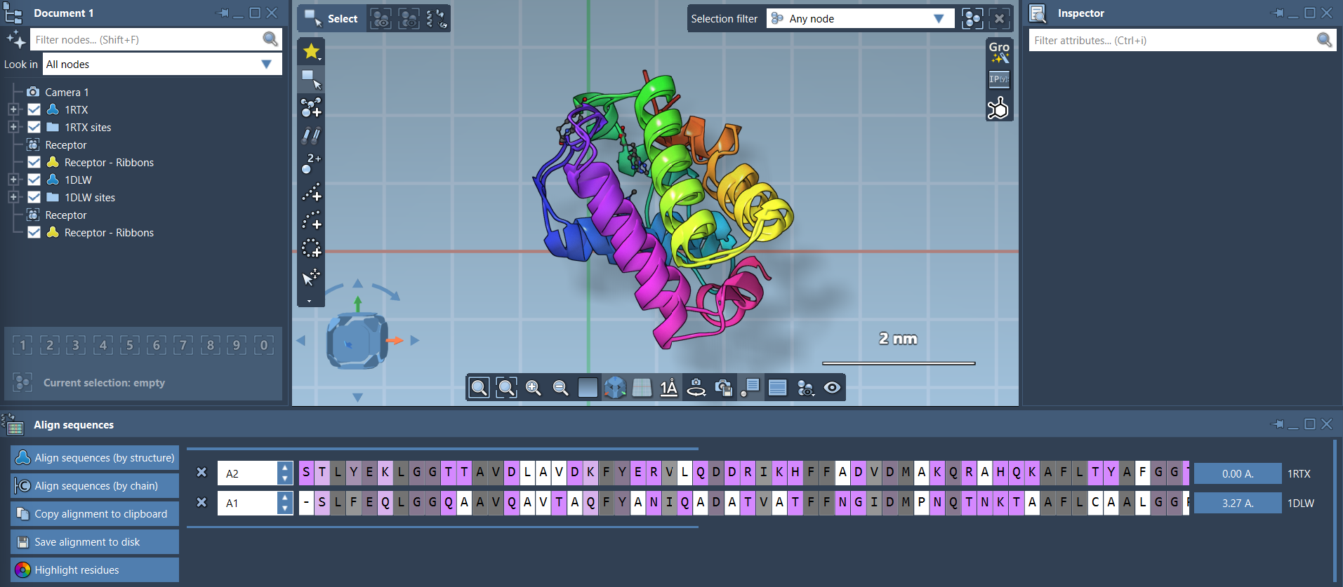
If the whole-protein fit does not answer your question, select the corresponding residues in both sequences and use the alignment button next to that selection. This superimposes the structures based only on the selected region. In the hemoglobin example, the first 20 residues are selected to focus on two similar alpha-helices that are not well aligned in the whole-protein view.

The practical lesson is simple: a global fit and a local fit answer different questions. Use the global fit to compare the overall structures; use a selected-region fit when you want to compare a specific structural feature. Keeping both views in mind helps prevent a single superposition from obscuring the region that matters to your analysis.
For secondary-structure ribbons, use Visualization > Visual model > Ribbons. Applying separate ribbon models to each protein lets you color them differently, and the Document view checkboxes can toggle atomistic views.
For the full workflow and additional examples, see the Protein Aligner documentation.
Note: SAMSON and all SAMSON Extensions are free for non-commercial use. Get SAMSON.