



Aligning protein structures is a routine task for molecular modelers, yet it can sometimes feel like solving a puzzle without clear guidance. Whether you’re tracking conserved residues, comparing mutants, or building homology models, structural alignment often paves the way for better protein insights. With SAMSON’s Protein Aligner, this process is integrated into an intuitive workflow that reduces complexity while enhancing precision.
Why Align Protein Structures?
If you’re working with protein models, you’ll likely need to understand how two or more structures compare. Protein alignments allow you to:
- Spot conserved residues that can shed light on functional sites or ligand binding.
- Detect conformational shifts across species or mutations.
- Contribute to accurate homology models for downstream molecular design efforts.
Getting Started
Before diving into the alignment, follow these preparatory steps:
- Clean the structures by removing extra solvent or ligands using the Protein Preparation & Validation extension.
- If needed, fetch example proteins (e.g., hemoglobin structures
1DLWand1RTX) via Home > Fetch. - Navigate to Home > Align to launch the Protein Aligner interface
 .
.
These steps ensure that your input data is clean and ready for meaningful comparisons.
Whole-Protein Structural Alignment
Once inside the Protein Aligner, you can align one protein structure to another using the built-in tools:
- Ensure no residues are selected in the structure (this tells the aligner to prioritize whole-structure superposition).
- Click the Align to this button next to the first model’s row. The other model is automatically superimposed, and the resulting root mean square deviation (RMSD) is displayed (e.g., 3.27 Å).
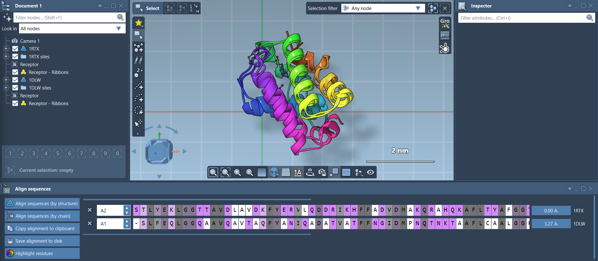
Overlaying the proteins makes it easier to visualize structural similarities or deviations and extract biologically relevant insights.
Region-Specific Alignment
What if you’re only interested in a specific part of the protein, such as a set of alpha-helices? SAMSON lets you dynamically align specific regions within a protein:
- Select the residues of interest directly from the Protein Aligner interface or through the viewport.
- Click the alignment button next to the selection to superimpose only the chosen residues.
This approach is invaluable when evaluating domain-specific interactions or flexible regions. For example, aligning the first 20 residues in two hemoglobins highlights subtle differences in the alpha-helices’ orientations.

Visualizing and Fine-Tuning Your Alignment
Enhance and streamline your structural analysis with a few additional visualization tips:
- Add secondary structure representations (e.g., ribbons) via Visualization > Visual model > Ribbons. Assign unique colors to each protein for clarity.
- Interacting with the alignment is easy—hover over residues to view their IDs, click one to highlight all aligned partners, or use keyboard shortcuts to refine your selection.
These tools help you focus on specific aspects of structural differences while maintaining context for the entire protein.
Next Steps
Once proteins are aligned, consider taking these additional steps:
- Export alignment data for building high-accuracy homology models.
- Use conserved residues to map ligand-binding sites for drug discovery.
- Repeat analyses with additional chains or mutations to widen the scope of insights.
Structural alignment isn’t just about overlaying proteins; it’s about extracting actionable knowledge—and with tools like Protein Aligner, this process becomes accessible and efficient.
To learn more about the Protein Aligner, visit the original documentation here.
Note: SAMSON and all SAMSON Extensions are free for non-commercial use. You can get SAMSON at https://www.samson-connect.net.