



Protein structure alignment is a routine task for molecular modelers, but ensuring accuracy while maintaining simplicity in workflows can be an ongoing challenge. If you’re comparing homologous proteins across species, monitoring conformational changes in mutants, or working on structural alignment for downstream applications, SAMSON’s Protein Aligner provides a practical and efficient solution. In this post, we’ll dive into how you can use the Protein Aligner extension in SAMSON to align protein structures in a few simple steps.
Understanding Protein Superposition
Aligning protein structures allows researchers to identify conserved residues, compare structural variations, and prepare homology models. However, partial matches, the presence of solvent/ligands, and complex selections often leave many unsure of how to streamline this process.
With SAMSON’s Protein Aligner, full-protein and region-specific alignments can be executed effortlessly, whether for whole proteins or individual structural features of interest. Let’s explore an example to better understand how to align structures effectively in SAMSON.
Step-by-Step Guide to Structural Alignment
Step 1: Load Your Proteins
In SAMSON, first fetch or load the proteins you wish to compare. For example, let’s align two hemoglobins: 1DLW and 1RTX. Head to Home > Fetch, enter the PDB codes 1DLW and 1RTX, and click the Load button to import the structures.

Step 2: Clean the Structures
If your structures include solvent molecules, ligands, or alternate locations, cleaning them simplifies the alignment process. Use the Protein Preparation & Validation extension (Home > Prepare) to strip unwanted elements before proceeding.
Step 3: Launch the Protein Aligner
Next, navigate to Home > Align and open the Protein Aligner extension. This tool allows you to align both sequences and 3D structures with a user-friendly interface. Let’s continue with aligning the overall protein structure.

Step 4: Align Structures
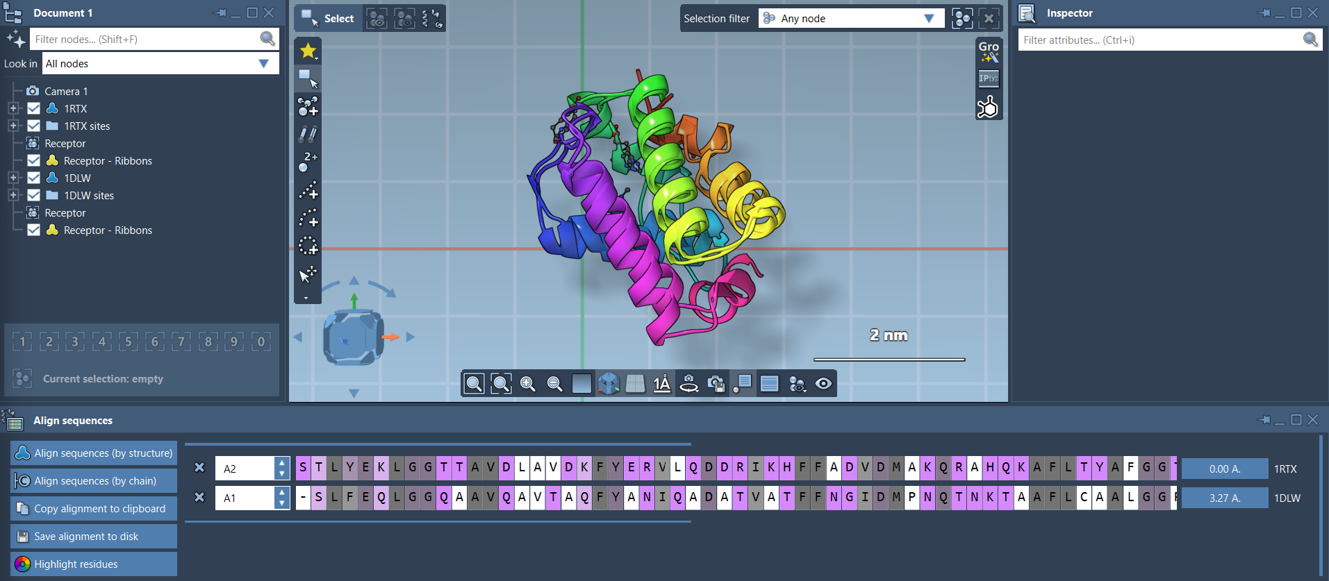
To perform a full-protein superposition, ensure no residues are selected and simply click Align to this on the row of your reference model. The aligned protein’s entry will then display its RMSD relative to the reference (e.g., 3.27 Å).
Upon alignment, the structures are superimposed for visual comparison. You can optionally enhance visual clarity by enabling Ribbons in Visualization > Visual model, with separate visual models applied to each protein for distinct coloring.
Step 5: Region-Specific Alignments
Sometimes, aligning the whole protein is less meaningful than focusing on a specific region, such as conserved domains or secondary structure elements. For instance, if you’re interested in aligning just the first 20 residues of both sequences, select these residues directly within the alignment view and click the alignment button (e.g., 0.0 Å).

You’ll achieve precise, region-based superposition. This is particularly useful for understanding localized structural differences or similarities.

Streamline Your Molecular Design Workflows
Whether you’re new to structure-based design or a seasoned researcher, SAMSON’s Protein Aligner lets you compare conformations, inspect conserved residues, and superimpose protein structures—all within the same interface. This saves time while improving the quality of insights in molecular modeling.
To explore the full documentation and learn more about SAMSON’s Protein Aligner, visit the official guide.
Note: SAMSON and all SAMSON Extensions are free for non-commercial use. To get started, download SAMSON at https://www.samson-connect.net.