



Protein modeling often requires comparing the structures of molecules to identify conserved residues, analyze conformations, or assist in downstream applications like homology modeling. However, aligning protein structures manually can be challenging and time-consuming—especially when precise superpositions are critical. SAMSON’s Protein Aligner Extension offers a convenient, streamlined solution to address this issue.
Why Use Protein Structure Alignment?
Aligning protein structures isn’t just about visualizing similarities—it provides real scientific advantages. For example, identifying conserved residues can help pinpoint critical regions for function or ligand binding. Comparing conformations across species or mutants is essential for understanding structural dynamics, and structural alignment serves as a foundation for creating reliable homology models.
Step-by-Step: Aligning Structures in SAMSON
With SAMSON’s intuitive interface, superimposing protein structures is straightforward. Let’s explore how to do it:
1. Preparing Your Proteins
Before starting the alignment process, it’s essential to load and clean your protein structures:
- Use Home > Fetch to retrieve proteins from public databases like PDB. For example, you could load hemoglobin structures
1DLWand1RTX. - If your structures contain unwanted solvent or ligands, clean them using the Protein Preparation & Validation tools in SAMSON.

2. Launching the Protein Aligner
Click on Home > Align to open the Protein Aligner interface:

3. Performing Whole-Protein Superposition
The simplest method to align your structures is to superimpose them as a whole:
- Ensure no residues are selected within the Protein Aligner.
- Choose the reference structure by clicking Align to this on the row corresponding to your first structural model.
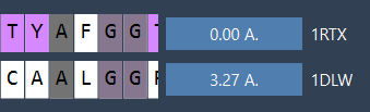
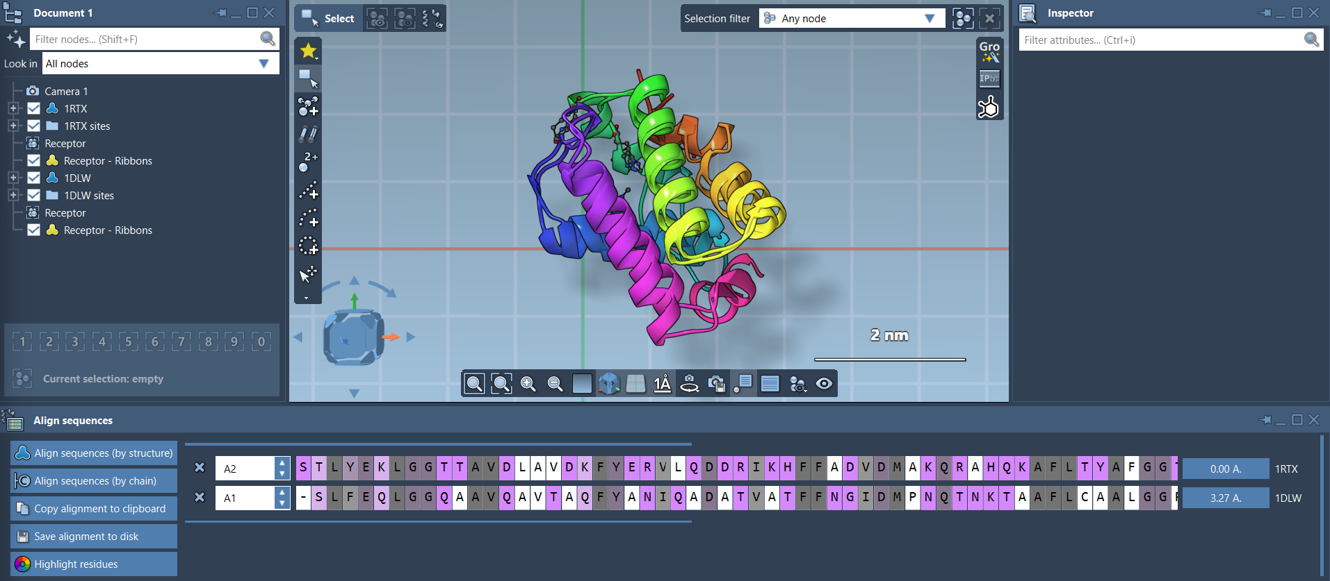
Once aligned, the second structure displays the Root Mean Square Deviation (RMSD), which quantifies the fit quality. For example, an RMSD value of 3.27 Å might be shown:
When the alignment is complete, the proteins are visually superimposed in the viewport:
4. Optional: Refining Alignment for Specific Regions
If you’re only interested in part of the structure (e.g., domains or secondary structure elements), you can align specific regions:
- Select the region of interest in both proteins within the Protein Aligner’s sequence view.
- Click the alignment button next to the selection to superimpose only that portion.
This tailored alignment can improve focus on features, such as conserved helices or binding-site residues.

Visual Enhancements
To improve the clarity of your aligned structures, consider adding visual aids:
- Enable Ribbons under Visualization > Visual model > Ribbons to represent secondary structures.
- Apply different colors to each protein using separate ribbon models for better differentiation. Simply select each structure before toggling visualization styles.
Next Steps and Applications
Once the structures are aligned, here are some suggested next steps:
- Export the alignment for use in homology modeling or molecular docking projects.
- Map conserved residues onto active or binding sites to guide drug design workflows.
- Explore conformations of additional proteins or mutants for further analyses.
Superimposing protein structures with SAMSON’s Protein Aligner Extension simplifies molecular comparison workflows while maintaining scientific rigor. To explore further capabilities and applications of the Protein Aligner, visit the full documentation at SAMSON Documentation.
SAMSON and all SAMSON Extensions are free for non-commercial use. You can get your copy at SAMSON Connect.