


Understanding viral proteins and their mechanisms is critical for researchers, especially molecular modelers, working on advancing virology, immunology, and drug design. One of the most intriguing examples is the SARS-CoV-2 spike protein, which plays a crucial role in the virus’s ability to infect human cells. How does it work? Let’s delve into its fascinating opening motion and why it matters in the fight against COVID-19.
The Key Role of the Spike Protein
The spike protein is a transmembrane protein that allows SARS-CoV-2 to attach to and enter human cells. Positioned on the virus surface, the spike enables infection by first binding to the ACE2 receptor on the cell membrane. This receptor-binding activity exposes the virus’s RNA, kickstarting the infection process.
The motion from the spike’s closed state to its open state is crucial as it allows the receptor-binding domain (RBD) to emerge and engage with the ACE2 receptor. Only the open state can successfully initiate this engagement, making it a key target for therapies and vaccine design.
Viewing the Spike: Structural Perspectives
The structure of this spike protein can be visualized in fascinating ways. Side and top views of the spike’s configurations reveal its complexity. Below are two important visualizations:

 The side view of the spike: Gaussian surface representation (left) vs. secondary structure representation (right).
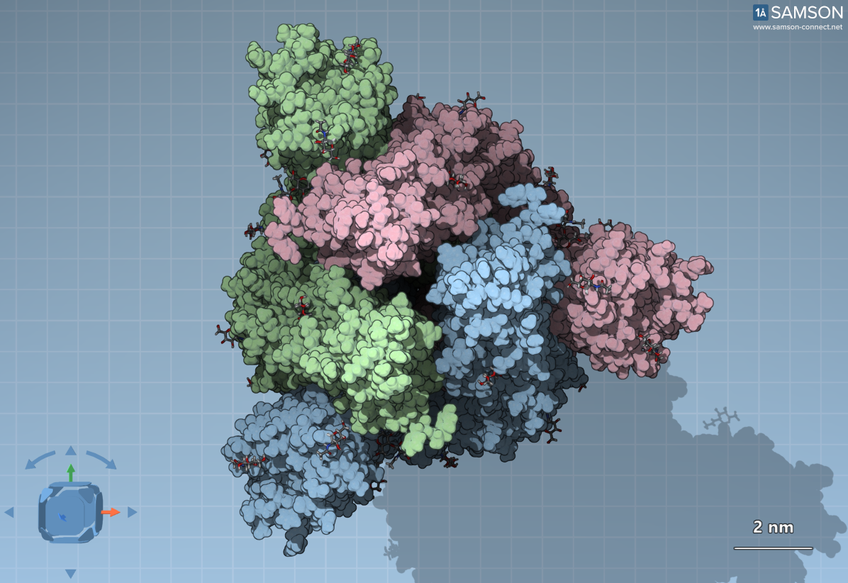
When viewed from the top, we see the C3 symmetry formed by three S-protein copies:
Why This Motion Matters to Researchers
The spike’s structural transitions are not merely academic curiosities—they directly inform therapeutic interventions. Understanding this motion helps modelers and researchers develop antibodies or inhibitors that target exposed regions during the opening process, minimizing virus-cell interactions.
A noteworthy challenge is that parts of the spike are disguised by sugar molecules to evade the human immune system. However, the receptor-binding domain, required for engaging ACE2 receptors, remains partly exposed. These exposed regions are often the focus of neutralizing antibodies.
Animation: Spike in Motion
What does the spike’s transition between its closed (down) state and open (up) state look like? These animations not only illustrate the process but also emphasize the dynamic nature of molecular interactions.
You can explore the computed trajectory files here to model the spike’s motion in your simulation environment.
Ready to Dive Deeper?
The motion of the SARS-CoV-2 spike provides invaluable insights for molecular modelers aiming to understand viral behavior or contribute to therapeutic development. To learn more about the role of the spike and how to visualize or simulate its transitions, visit the detailed documentation at this link.
Note: SAMSON and all SAMSON Extensions are free for non-commercial use. You can download SAMSON at https://www.samson-connect.net.