


When modeling protein-ligand complexes from NMR data, one of the trickiest steps is translating NOESY cross-peaks into distance restraints that can be used for structure prediction. In particular, if you’re using NMR2 in SAMSON to interpret methyl-based NOESY data, you’ll encounter a simple but essential interface where all your distance information must be entered manually.
What’s unexpectedly tedious for many molecular modelers is ensuring that the syntax is exactly as expected, learning what qualifies as a valid site name (pseudo-atom? methyl group?), and understanding how distance restraints affect the calculation process downstream.
Format overview
In the NMR2 extension, distance restraints must follow this format:
|
1 |
SITE_1 SITE_2 = LOWER_BOUND UPPER_BOUND |
For example:
|
1 |
Q4 M5 = 2.18 3.88 |
Here, Q4 is a pseudo-atom in the ligand, M5 is an unassigned methyl group, and the restraint implies that these two sites should remain between 2.18 and 3.88 angstroms apart.
When manual input becomes practical
Even though the restraints must be typed or pasted manually, it’s possible to prepare them efficiently. A typical list might look like this:
|
1 2 3 4 5 6 7 8 9 10 11 12 13 14 |
Q4 M5 = 2.18 3.88 H8 M5 = 3.80 5.70 Q4 M1 = 3.73 6.64 H7 M3 = 4.42 6.62 H5 M2 = 2.36 3.54 Q M3 = 2.88 5.12 H7 M4 = 2.91 4.36 H7 M1 = 4.63 6.94 H5 M4 = 2.17 3.25 Q4 M4 = 2.81 4.99 H8 M4 = 3.92 5.88 H7 M5 = 3.56 5.34 Q4 M2 = 3.52 6.25 H8 M1 = 3.58 5.36 |
✅ Tip: Copy this list as a block into the Distances field in the NMR2 interface.
Each line defines a NOE-based restraint, and using this format allows NMR2 to evaluate how well a predicted structure complies with available NOESY data.
What actually happens behind the scenes?
Once you provide these restraints and run a calculation, NMR2 searches over possible methyl assignments and ligand poses that satisfy your distance information. The software computes combinations and ranks them by how well the restraints are met (using a target function), and how much steric clash is avoided (using a Van der Waals function).
This part is fully automated. But it relies heavily on the accuracy and completeness of your input restraints. Poorly formatted entries or inconsistency in site naming can lead to uninformative results or errors.
Identifying pseudo-atoms and methyls
To define the correct site names (like Q, M4, or H7), you first need to:
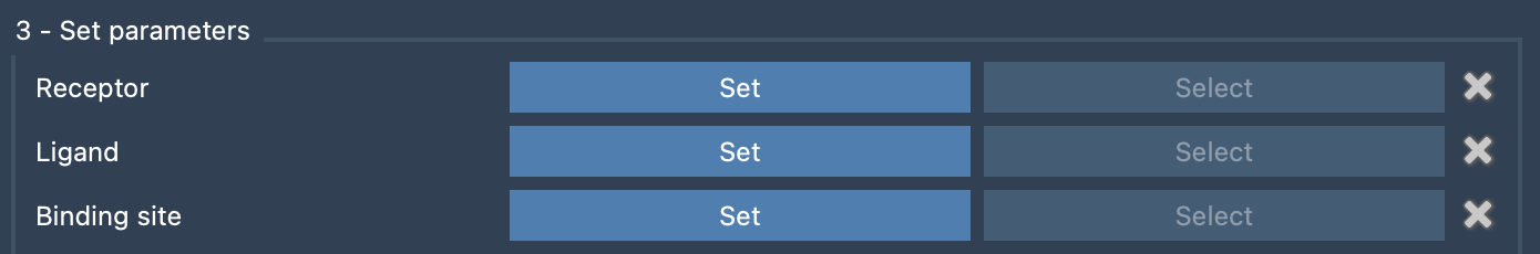
- Set the protein and ligand components in the NMR2 interface.
- Let NMR2 automatically generate pseudo-atoms for the ligand when selected.
- Use visual inspection to cross-check names, or select them from the drop-downs if possible.
These names must exactly match the labels used in your restraints. It helps to open atom name labels in SAMSON visually to keep tabs on available identifiers.
Wrapping up
Manual distance restraint input may look like a mundane step in your modeling workflow—but it’s a crucial one. Getting it right once ensures smooth execution of the NMR2 protocol, and more meaningful ligand placement predictions.
To dive deeper into the full workflow—including system setup, assigned methyls, side-chain flexibility, and analyzing results—you can read the full guide here.
SAMSON and all SAMSON Extensions are free for non-commercial use. You can download SAMSON at https://www.samson-connect.net.